About Us

The Immunomonitoring Laboratory (IML) provides cutting-edge technology platforms for the comprehensive immunophenotyping of blood and tissues. Our services allow investigators to monitor the impact of therapeutic interventions on immunologic profiles, interrogate mechanisms and pathways related to treatment protocols and advance the conceptual basis for targeted treatment strategies. We collaborate with scientists, clinicians and industry partners involved in clinical trials and discovery-based immunologic research.
Interested in learning more? Contact us today at [email protected].
Our Team


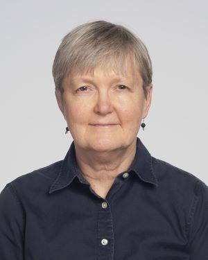
Jennifer Ko, MD, PhD
Medical Director, Immunomonitoring Lab


Technologies
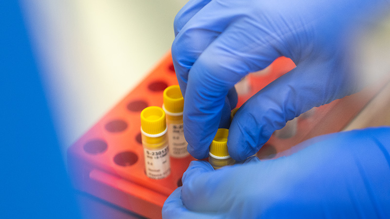
Sample Acquisition, Processing, and Banking
The IML offers cost-effective standardized and optimized procedures for blood and tumor tissue processing. We continuously monitor our procedures to ensure consistent yield of viable cell products. We also offer assistance with paraffin tissue block selection and retrieval, as well as assay-specific processing. In collaboration with the Pathology and Laboratory Medicine Central Biorepository, the IML also offers acquisition, barcoding, and controlled storage of procured samples and/or cell products. We utilize the scientific data management system LabVantage® for biospecimen annotation, report generation, and quality control for ensured compliance with regulatory standards.
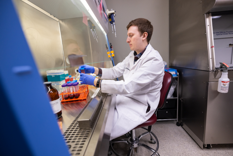
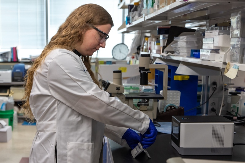
Flow Cytometry
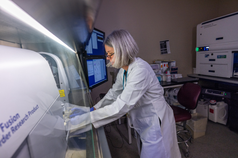
The IML offers high-parameter flow cytometry using the BD FACSymphony®A5 and four-way cell sorting using the BD FACSAria™ Fusion, both of which have five lasers (355nm, 407nm, 488nm, 532nm, and 633nm). The Symphony allows interrogation of up to 29 parameters, not including forward scatter and side scatter. The Fusion, located in a Baker biosafety cabinet, allows up to 20 parameters and can perform bulk sorting into tubes or single cell sorting in plate wells.
Single-Cell Immune Profiling
10x Genomics
The 10x Genomics Single-Cell Immune Profiling platform allows for the multimodal interrogation of gene expression, paired-chain TCR sequences, BCR sequences, and cell surface protein expression at single cell resolution. Single cell suspension can be prepared in your lab or added onto existing biobanking services run through the IML. Cell surface protein detection is highly customizable – design your own panel using any BioLegend TotalSeq™-C antibodies or use a universal 130 antibody cocktail optimized for immune cell characterization. Experiments can be run with all four modalities simultaneously or with any combination of the four.
Parse Biosciences
The Parse Biosciences Evercode Whole Transcriptome kit harnesses the power of combinatorial barcoding to run cost-effective, ultra-high-throughput single cell RNA-seq experiments. Up to 1 million cells split across 1-96 samples can be analyzed simultaneously, and both gene expression and TCR sequencing are available. The Parse Evercode platform uses fresh cells as input, allowing for greater flexibility when planning and running single cell experiments. Cells can be fixed immediately after sample collection and stored in the freezer for up to six months. Researchers can choose to fix their own samples or add cell fixation onto biobanking services run through the IML.
Liquid Biopsy
Personal Genome Diagnostics (PGDx)
The IML offers in-house circulating tumor DNA sequencing in partnership with PGDx using their Elio Plasma Complete platform. This assay examines 521 pan-cancer related genes and provides sequence mutations, insertions and deletions, copy number variations, blood tumor mutational burden, microsatellite instability, and select amplifications and translocations all from a single blood draw.
Spatial Transcriptomics
10X Genomics - Visium
Spatially resolved transcriptomic technologies allow investigators to gain novel insight into tissue biology. The ability to measure gene expression with spatial context yields valuable information about the organization of cell types and states in tissue, which furthers understanding of the complexity of their interactions and relationships.
The IML offers spatial transcriptomic analysis using the 10X Genomics Visium Spatial Gene Expression platform. Visium is a next-generation molecular profiling solution for analyzing total mRNA expression that maps the whole transcriptome with morphological context in entire FFPE as well as fresh frozen tissue sections.
Proteomics
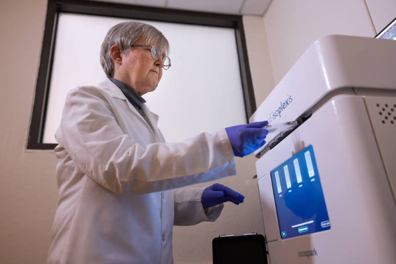
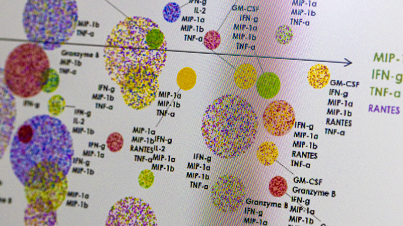
Bruker Cellular Analysis (Isoplexis)
The IML's IsoSpark instrument, which runs the IsoCode and CodePlex Chips, is a versatile, precision-automated platform designed to understand and characterize differences among immune cells by mapping thousands of cells per sample.
IsoCode's single-cell secretome solution allows for complete single-cell functional characterization. It will detect rare subsets of highly functionally active cells to reveal potential biomarkers of response as well as functional biological drivers of persistence and potency.
CodePlex Chip immunoassays allow automated multiplex proteomics analysis in very low sample volume. They offer a faster and more streamlined approach to generating multiplexed bulk cytokine data via a fully automated workflow.
Olink
The IML's Olink Signature Q100 is a dedicated system specifically designed for the readout of Olink Target (96 & 48-plex), Olink Flex (15–21 chosen assays) and Olink Focus (custom design) protein biomarker panels. These are focused on specific disease areas or biological processes and offer Proximity Extension Assay (PEA) technology coupled with a qPCR readout. The disease- or biology-focused Olink Target 96 & 48 panels enable rapid analysis with minimal sample consumption and exceptional data quality, with protein concentrations reported in standard units (pg/mL) as default and optionally in relative (NPX) units. Using just 1 µl of sample, up to 92 proteins with Target 96 and 45 proteins with Target 48 are assayed simultaneously on each panel, and results are generated within 24 hours.
Highlighted Studies
Kidney Cancer
- CASE 5816: A Single-arm Phase II Trial of Intermittent Nivolumab in Patients with Metastatic Renal Cell Carcinoma who Have Received Prior Anti-Angiogenic Therapy
- CASE 12815: A Phase Ib Trial of Neoadjuvant Durvalumab (MEDI4736) +/- Tremelimumab in Locally Advanced Renal Cell Carcinoma
Bladder Cancer
- CASE 6819 A Phase II Study of Intermittent Checkpoint Inhibitor Therapy in Patients with Advanced Urothelial Carcinoma
Brain Cancer
- CASE 1317: A Randomized Phase 2 Open Label Study of Nivolumab plus standard dose Bevacizumab versus Nivolumab plus low dose Bevacizumab in Recurrent Glioblastoma (GBM)
- CASE 2317: Phase I Study of Ibrutinib with Radiation and Temozolomide in Patients with Newly Diagnosed Glioblastoma
- CASE 3317: Phase I Study of Ruxolitinib with Radiation and Temozolomide in Patients with Newly Diagnosed Grade III Gliomas and Glioblastoma
- CASE 7318: Regorafenib in Bevacizumab Refractory Recurrent Glioblastoma
- CASE 4316: Expedited Laser Interstitial Thermal Therapy and Chemoradiation for Patients with Newly Diagnosed High Grade Gliomas
Breast Cancer
- CASE 3113: Overcoming Stromal-specific Immune Escape Mechanisms by Targeted Immunotherapy.
- CASE 8119:Evaluation of immunologic and serologic biomarkers for prediction of response to checkpoint antibody therapy in metastatic triple negative breast cancer (TNBC)
Melanoma
- CASE 1617: Germline Mutations in High Risk Melanoma Patients
- CASE 1619: Leveraging ctDNA Analysis to Improve Early Detection of Cancer Recurrence in the High-Risk Adjuvant Melanoma Setting
- CASE 2619: Phase II Trial of Nivolumab in Combination with Talazoparib in Patients with Unresectable or Metastatic Melanoma with mutations in BRCA or BRCA-ness
- CASE 6620: Low-Dose-Rate Brachytherapy Combined with Immune Checkpoint Inhibition in Cancer
Publications
Alban TJ, Grabowski MM, Otvos B, Bayik D, Wang W, Zalavadia AH, Makarav V, Troike KM, McGraw M, Rabljenovic A, Lauko A, Neumann CK, Roversi G, Waite KA, Cioffi G, Patil N, Tran TT, McCortney K, Steffens A, Diaz-Montero CM, Brown JM, Egan KM, Horbinski C, Barnholtz-Sloan JS, Rajappa P, Vogelbaum MA, Bucala R, Chan TA, Ahluwalia MS, Lathia JD. The SNP rs755622 is associated with immune activation in glioblastoma [published online ahead of print, 2023 May 30]. JCI Insight. 2023;e160024. doi:10.1172/jci.insight.160024
Pavicic PG Jr, Rayman PA, Swaidani S, Rupani A, Makarov V, Tannenbaum CS, Edwards RP, Vlad AM, Diaz-Montero CM, Mahdi H. Immunotherapy with IL12 and PD1/CTLA4 inhibition is effective in advanced ovarian cancer and associates with reversal of myeloid cell-induced immunosuppression. Oncoimmunology. 2023;12(1):2198185. Published 2023 Apr 10. doi:10.1080/2162402X.2023.2198185
Li C, Phoon YP, Karlinsey K, Tian YF, Thapaliya S, Thongkum A, Qu L, Matz AJ, Cameron M, Cameron C, Menoret A, Funchain P, Song JM, Diaz-Montero CM, Tamilselvan B, Golden JB, Cartwright M, Rodriguez A, Bonin C, Vella A, Zhou B, Gastman BR. A high OXPHOS CD8 T cell subset is predictive of immunotherapy resistance in melanoma patients. J Exp Med. 2022;219(1):e20202084. doi:10.1084/jem.20202084
Roopkumar J, Swaidani S, Kim AS, Thapa B, Gervaso L, Hobbs BP, Wei W, Alban TJ, Funchain P, Kundu S, Sangwan N, Rayman P, Pavicic PG Jr, Diaz-Montero CM, Barnard J, McCrae KR, Khorana AA. Increased Incidence of Venous Thromboembolism with Cancer Immunotherapy. Med. 2021;2(4):423-434. doi:10.1016/j.medj.2021.02.002
Tannenbaum CS, Rayman PA, Pavicic PG, Kim JS, Wei W, Polefko A, Wallace W, Rini BI, Morris-Stiff G, Allende DS, Hamilton T, Finke JH, Diaz-Montero CM. Mediators of Inflammation-Driven Expansion, Trafficking, and Function of Tumor-Infiltrating MDSCs. Cancer Immunol Res. 2019;7(10):1687-1699. doi:10.1158/2326-6066.CIR-18-0578
Lin L, Rayman P, Pavicic PG Jr, Tannenbaum C, Hamilton T, Montero A, Ko J, Gastman B, Finke J, Ernstoff M, Diaz-Montero CM. Ex vivo conditioning with IL-12 protects tumor-infiltrating CD8+ T cells from negative regulation by local IFN-γ. Cancer Immunol Immunother. 2019;68(3):395-405. doi:10.1007/s00262-018-2280-3
Pfannenstiel LW, McNeilly C, Xiang C, Kang K, Diaz-Montero CM, Yu JS, Gastman BR. Combination PD-1 blockade and irradiation of brain metastasis induces an effective abscopal effect in melanoma. Oncoimmunology. 2018;8(1):e1507669. Published 2018 Oct 11. doi:10.1080/2162402X.2018.1507669
Ko JS, Gastman BR, Conic R, Tellez Diaz Trujillo A, Diaz-Montero CM, Billings SD, Tarhini A, Funchain P, Atanaskova Mesinkovska N. Decreased T-Cell Programmed Death Receptor-1 Expression in Pregnancy-Associated Melanoma. Am J Dermatopathol. 2019;41(3):180-187. doi:10.1097/DAD.0000000000001286
Najjar YG, Rayman P, Jia X, Pavicic PG Jr, Rini BI, Tannenbaum C, Ko J, Haywood S, Cohen P, Hamilton T, Diaz-Montero CM, Finke J. Myeloid-Derived Suppressor Cell Subset Accumulation in Renal Cell Carcinoma Parenchyma Is Associated with Intratumoral Expression of IL1β, IL8, CXCL5, and Mip-1α. Clin Cancer Res. 2017;23(9):2346-2355. doi:10.1158/1078-0432.CCR-15-1823