About Us
The Flow Cytometry Core provides investigators with a resource for quantitative studies of cells, extracellular vesicles, and single-cell gene transcriptome analyses. The Core offers sample acquisition and analysis, cell sorting, and consultation for experimental design, interpretation and troubleshooting.
Radioactive materials are prohibited.
To recognize the Flow Cytometry Core personnel or technology for support of your research, please refer to the Acknowledgment tab.
Contacts
Kewal Asosingh, PhD, SCYM(ASCP)
Staff
Board Certified Specialist in Cytometry
[email protected]
Lab Profile
Eric Schultz, SCYM (ASCP)
Shared Laboratory Resource Specialist
Services
Capabilities:
- Sony ID7000 spectral flow cytometer with advanced high-parameter flow cytometry capabilities. The instrument has six lasers (320nm, 355nm, 405nm, 488nm, 561nm, 637nm) and 182 detectors for complete spectral data acquisition, allowing +45 color panels, load-and-walkaway data acquisition and advanced fluorescence subtraction tool. The ID7000 is ideal for analyzing samples with complex heterogeneous cell populations.
- Cytek Aurora spectral flow cytometer equipped with five lasers (355nm, 405nm, 488nm, 561nm and 640nm excitation wavelengths), three scattering channels, 64 fluorescence channels and can acquire +45 color panels. Samples can easily be acquired from 96-well plate format or single tube mode. The Cytek Aurora is also equipped with an Enhanced Small Particle Detector (size limit 80nm) for extracellular vesicle studies.
- BD FACSymphony A5 SE versatile conventional/spectral cell analyzer housing five lasers (355nm, 402nm, 488nm, 561nm, 640nm) and 50 detectors.
- Becton-Dickinson LSRFortessa capable of 18 color and 20 parameter acquisition with 5 lasers (355nm, 407nm, 488nm, 561nm, and 641nm).
- Bigfoot High Speed Cell Sorter equipped with 48 fluorescence detectors, and five light scatter detectors offers six-way BSL2 cell sorting at high-speed (>70,000 events per second). This instrument can also sort small particles (>200nm) and is equipped with depolarized light scatter detectors for antibody-free gating of birefringent eosinophils and other cells.
- Becton-Dickinson SORP FACSAria Fusion high performance with ACDU, fixed alignment gel-coupled cuvette cell sorter. The BD SORP FACSAria Fusion cell sorter consists of 355nm, 405nm, 488nm, 561nm, and 637nm lasers, providing 20 parameters.
- MA900 cell sorter detects up to 14 parameters. Four excitation lasers (405, 488, 561, and 638 nm) are available on two beam spots. Fluorescence signals from each beam spot can be detected using free-form PMTs, which allows for the detection of up to 12 fluorescence parameters and two scatter parameters.
- All cell sorters are housed in specialty type Class II biological safety cabinets designed to offer personnel, product, and environmental protection from potentially aerosolized biological hazards.
- Zetaview QUATT Nanoparticle Tracker Analyzer.
- 10x Chromium Controller for single cell partitioning for transcriptome analysis.
- CellDrop Automated Cell Counter offers objective evaluation of cell counts, viability, aggregates and cell debris.
The Core provides data analysis with the following software:
- FlowJo (WatersTM Biosciences)
- ModFit LT (Verity)
- Sony ID7000 Software Sony
- terraFlow
- ZetaSphere
SpectroFlo, FACSDiva and Sasquatch (SQS) are for acquisition only.
Analysis workstations and software training are available for researchers.
Consultation:
All investigators are encouraged to meet with the Scientific Director of the Flow Cytometry SLR to discuss their experiment prior to ordering antibodies and other reagents. This meeting is often useful in that many potential problems can be averted before critical resources are used. SLR specialists are available for assistance with sample acquisition, analysis and presentation. Materials available for reference in the Flow Cytometry SLR include introductory tutorials available in iLab, several textbooks on the basics of flow cytometry and the current protocols in cytometry. In addition, the SLR frequently organizes training seminars for various levels of expertise. A meeting with both Flow Cytometry and Genomics SLR directors is mandatory for pilot 10x experiments. User consultations are free of charge!
Flow Cytometry Applications:
- Free of charge guidance in sample preparation and panel design, and affordable prices for instrument training and data analysis.
- Functional assays utilizing fluorescent probes such as quantification of mitochondrial organelle function, calcium influx and apoptosis, and quantification of expressible fluorescent proteins
- + 45 color cell surface and/or intracellular (cytokine) immunophenotyping.
- Measurements of soluble factors in biological fluids and cell culture supernatants using bead-based multiplex assays.
- Sample preparation and single cell partitioning using the 10x Chromium X.
- Six-way sterile electrostatic cell sorting of preclinical and clinical samples for functional studies.
- Extracellular vesicle enumeration and characterization.
SpectroFlo® is used on the Cytek Aurora for data acquisition. Sony ID7000 software is used on the ID7000 spectral analyzer. BD FACSDiva software is used to acquire data on the Fortessa, Fusion and Symphony A5 SE. Data can be imported/exported as FCS 2.0, 3.0, or 3.1 when using FACSDiva. List mode acquisition and storage of data allows for analysis and presentation using many different formats according to the needs of the investigator. Data files are permanently stored on Cleveland Clinic Research servers in lab folders, where they are easily accessible to all Cleveland Clinic Research employees with proper login accounts. FlowJo and ModFit software are available on Flow Cytometry SLR workstations for analysis. FlowJo Portal licenses can be purchased individually by contacting the Flow Cytometry SLR.
Getting Started with the Flow Cytometry Core:
Please visit our iLab page to schedule services. Users can find introductory materials, best practices and policies on our Links & Resources page.
Equipment
Sony ID7000 spectral flow cytometer with advanced high-parameter flow cytometry capabilities. The instrument has six lasers (320nm 20 mW, 355nm 50 mW, 405nm 100mW, 488nm 150mW, 561nm 100mW, and 637nm 140mW) and 182 detectors for complete spectral data acquisition, allowing up to +45 color panels or more, limited only by the fluorochromes available. Load-and-walkaway data acquisition is provided by automated sampling. Another distinguishing feature of this equipment is the ability to easily subtract autofluorescence from multiple subsets to reduce background signal. The ID7000 is ideal for analyzing samples with heterogeneous cell populations, such as tumors and organs, because it detects dim and rare populations with the highest sensitivity available today. Regardless of the complexity of the panel, the overall workflow of the instrument is very simple. The use of this instrument must be acknowledged in manuscripts by adding “This work utilized a Sony ID7000 that was purchased with funding from the National Institutes of Health SIFAR grant S10OD025207.”
CytekTM Aurora spectral cell analyzer housed in the Flow Cytometry Core is equipped with five lasers (355nm: 20mW; 405nm:100mW; 488nm:50mW; 561nm:50mW; 640nm:80mW). Spectral flow cytometry technology has propelled over the past years and offers several key advantages compared to conventional flow cytometry. Traditional flow cytometry samples a narrow band of the optical spectrum. While this is very useful and has been the standard in flow cytometry over the past decades, spectral flow cytometry provides the full fluorescence emission spectrum measured on each cell, enabling easy separation of complex mixtures of fluorochrome combinations that are challenging to co-detect using traditional flow cytometry. The Aurora utilizes avalanche photodiodes resulting in higher resolution, sensitivity, and linear signals compared to conventional systems. The optical configuration is filterless, making the instrument user-friendlier and eliminates errors caused by potential wrong settings. A short-wavelength (violet laser) side scatter detector makes enumeration and immunophenotyping of microparticles possible on the Aurora. Vacuum fluidics allow quantification of absolute numbers. An autofluorescence extraction function allows better resolution of dim signals by subtraction of high autofluorescence that is inherent to some cells such as macrophages, epithelial and endothelial cells. The overall workflow of the instrument is straightforward, regardless of the complexity of the panel.
This cell analyzer is capable of 48 colors or 50 parameter acquisition with five lasers (349nm-60mW; 405nm-200mW; 488nm-150mW; 561nm-150mW and 637nm-140mW).
- GaAsP (Gallium, Arsenide, Phosphide) PMTs reduce noise and increase sensitivity in red and near-infrared emission channels.
- This instrument has some spectral capabilities. Our users are recommended to utilize the Cytek Aurora or ID7000 for spectral flow cytometry.
A BD LSR Fortessa capable of 18 colors or 20 parameter acquisition with five laser lines including: 355nm, 407nm, 488nm, 561nm, and 641nm. This cytometer is dedicated for emergency use or same day (less than 24 hours) experiments by trained users.
Fortessa Configuration(View PDF)
The BigFoot is a high-speed cell sorter, 53 parameter cell sorter equipped with 349nm-100mW, 405nm-100mW, 488nm-125mW, 561nm-120mW and 640nm-100mW lasers.
- 48 fluorescence detectors and 5 light scatter detectors.
- Straight-down, 2-way, 4-way and 6-way BSL2 cell sorting at high-speed (>70,000 events per second).
- Small particle sorting (>200nm).
- Depolarized light scatter detectors for antibody-free gating of birefringent eosinophils and other cells.
- Sample input: 6 tube input positions for 1.5 mL, 5 mL, or 15 mL tubes; temperature control (4–37°C) and agitation for all positions.
- Sort output: Up to six-way sorting into tubes; configurable tube holders include 1.5 mL, 5 mL, 15 mL, and 50 mL adapters. Multiway microwell plate sorting up to 1,536 wells. Temperature control (4–37°C) for all media types.
- Nozzle: 70µm, 100µm and 120µm ceramic nozzle tips with adjustable pressure settings.
- Biocontainment: Integrated Class II biocontainment cabinet for protection of sample and user. Separate AES for sort chamber evacuation.
- This instrument has some spectral capabilities as well.
The BD SORP FACSAria Fusion, 20 parameter, 4-way cell sorter is equipped with 355nm, 405nm, 488nm, 561nm, and 637nm laser lines. The sorter is enclosed in a Class II Type A2 biosafety cabinet (BSC), designed to offer personnel, product, and environmental protection from potentially aerosolized biological hazards. This cell sorter has a capability of up to 4-way bulk sorting, single-cell plate index sorting, multi-functional temperature control for sample injection and collection.
Additional options included:
- 70, 85, and 100-micron nozzles
- Two- and four-way sorting into microtubes, 12x75-mm tubes
- Two-way sorting into 15 mL tubes
- Automated Cell Deposition Unit (ACDU) allows for multi-well plate and slide sorting
- Single-cell index plate sorting
- Accepts various sizes of sample loading tubes
- Temperature controlled chamber for sample loading (RT, 4oC, 37oC)
- Temperature control system for chilled (5oC) collection devices (tubes and multi-well plates)
Our first cell sorter that users may be trained to operate as self-use! This easy to operate cell sorter detects up to 14 parameters. Four excitation lasers (405nm-120mW, 488nm-80mW, 561nm-110mW, 638nm-110mW) are available on 2 beam spots. Fluorescence signals from each beam spot are detected using free-form PMTs.
- 12 fluorescence detectors and 2 light scatter detectors.
- One-way (plate) 2-way, 4-way BSL2 cell sorting.
- Sample input: 0.5-mL, 1.5-mL, 5-mL, and 15-mL tubes; temperature control (5 or 37°C) and agitation for all positions.
- Sort output: 6-, 12-, 24-, 48-, 96-, and 8-, 96- or 384-well PCR plates. Temperature control (5 or 22.5°C).
- Sorting chip size: 70µm, 100µm and 130µm.
- Biocontainment: custom class A2 level II biosafety cabinet for protection for personnel and cells. The cabinet has an integrated aerosol management system that operates autonomously to actively evacuate aerosols from the sort collection chamber. Aerosol evacuation via two routes maximizes protection.
View MA900 configuration (PDF).
The ZetaView is a nanoparticle tracking analyzer to determine the size histogram and concentration of exosomes and microparticles in biological fluids.
- Detection range 20nm-2,000nm.
- Zeta potential measurement.
- Measurement of particle size at single particle level using light scattering and fluorescence.
- Ability to co-localize the fluorescence signal and the scattering signal at the single particle level (2 parameters at a time) in order to identify the precise subpopulation that carries the fluorescent label.
- Temperature controlled flow cell.
A Chromium X 10x Genomics instrument is available for automated single cell preparations and single cell gene expression applications. Evaluation of the single cell sample preparation, cell sorting (if needed), GEM creation, thermal cycling and cDNA amplification are performed in the Flow Cytometry SLR. Further amplification and clean-up of libraries and sequencing are performed in our Genomics SLR.
Eliminates the need for disposable plastic slides with CellDrop's direct-to-sample technology. Accurately quantify cells across a wide range of densities by adjusting chamber height to accommodate various sample types. A reliable and low-waste solution for routine and high-throughput workflows.
- Cells/mL, cell size and % viability reported.
- Cell viability analysis in the presence of debris using acridine orange (AO) for identification of nucleated cells and propidium iodide (PI) for cell viability.
- Dynamic range 7 x 10^2 – 2.5 x 10^7 cells/mL.
- Sample volume: 5µL, 10µL and 40µL.
- Cell size range 4 – 400 µm.
Links & Resources
Current Protocols in Cytometry is available online through the Cleveland Clinic Alumni Library. This resource is reached by searching the library catalog, in the A-Z list by resource name, or in Find Journals.
- Red Blood Cell Lysis Buffer (View PDF)
- Antibody Titration Basics (View PDF)
- Immunophenotyping Methods and Protocols (View PDF)
- 10 Simple Clicks to Schedule Time on the Flow Cytometer (Download Word File)
- Flow Cytometry Core iLab Guide (View PDF)
- Access the Lerner Flow Cytometry Core (CWRU) (View PDF)
- Cell Sorting Procedure (Download Word File)
- FlowJo Portal Licensing Policy (View PDF)
- FlowJo Portal User Instructions (View PDF)
- Detection of Intracellular Cytokines by Flow Cytometry (View PDF)
- Relevance of Antibody Validation and Titration for Flow Cytometry (View PDF)
Links
- The Purdue University Cytometry Laboratories maintain valuable email discussions on flow topics from researchers around the world.
- Visit Washington State University's Taxonomic Key Program website for comparative analysis of the specificity of mAbs specific for leukocyte differentiation molecules intra- and cross species.
- Thermo Fisher Scientific's Fluorescence SpectraViewer provides a viewer for selecting fluorescent markers and assessing spillover estimates.
- FluoroFinder is an alternative fluorescence spectrum viewer.
- Great Lakes International Imaging and Flow Cytometry Association (GLIIFCA) holds an annual meeting in the fall of each year and sponsors this website to serve as a clearing house for information of interest to researchers in the Great Lakes region.
- International Society for Advancement of Cytometry (ISAC) provides a forum for exchanging information related to the utilization of analytical cytology methods.
- Bio-Rad provides an Introduction to Flow Cytometry-Basics Guide.
- Visit the FlowJo website for Tutorials, Tech Notes and Demo Data.
- BD Biosciences provides an overview of BD reagents and protocols including fluorochrome conjugated antibodies, buffers, and kits for intracellular staining of cytokines, phosphoproteins, and transcription factors.
- Life Technologies’ Flow Cytometry Resource Center offers free basic flow cytometry tutorials and educational webinar series, plus tips and tricks to optimize your flow cytometry experiments.
- Learn more about SegQeq, FlowJo’s bioinformatics platform .
- BenchSci is an AI-assisted reagent selection.
- Visit the International Society for Advancement of Cytometry's BioSafety Standards webpage for biosafety guidelines.
Frequently Asked Questions
- Can I sort unfixed human samples?
Yes. A BioProtect III Walk-In Clean Air and Containment Cabinet houses the FACSAria II. The BioProtect III is a specialty type Class II biological safety enclosure designed to offer personnel, product, and environmental protection from potentially aerosolized biological hazards. When a sort has a biohazard potential, a biohazard form must be completed prior to each sort via iLab.
- Can I run fluorochrome X with fluorochrome Y?
To determine the spectral overlap of varied fluorochromes visit BD Biosciences or FluoroFinder. Select fluorochromes from the list and compare their excitation and emission wavelengths to get an idea of compatibility. Fluorochromes whose emission peaks overlap completely may not be used together.
- How many cells do I need for each tube? What should the volume be?
Analytical experiments, under ideal circumstances, one million cells per tube are standard. In many situations, few cells may be available for each of the controls and experimental samples. The minimum number of cells required is going to vary based on needs. For very simple experiments, tens of thousands of cells may be needed. Multicolor experiments that subset out small percentages of cells may require several million cells per tube in order to acquire appropriate numbers of the target populations. (ref: Cytometry A. 2008 May;73(5):384-5. Roederer, M. How many events is enough?) A minimum of 200ul volume in each sample tube is required.
- What controls do I need for my experiment?
Instrument controls consist of samples needed to compensate/unmix the different fluorochromes and to choose starting baseline voltages if optimal PMT voltages are not used. To properly compensate/unmix for an experiment, an unstained control (cells with NO fluorochromes) and a single color control for each fluorochrome for that experiment are needed. The instrument is set up using these controls for compensation/spectral unmixing. These controls are mandatory. FMO controls are highly recommended to set gate boundaries correctly. A consultutation with the Core Director is highly recommended to determine application specific controls.
- Do I really need all these controls?
Yes. At times, the Flow Cytometry Core is called upon to setup the instruments without these controls. In these circumstances it is the Core's policy to inform the investigator that the instrument is being set so the data looks a certain way based on expectations and experience. Results from these experiments are invalid and need to be repeated with the proper controls. Any conclusions drawn under these circumstances are highly suspect at best.
- Do I really need to run compensation controls everyday?
Yes. Even though the instruments are QC'd on a daily basis to measure stability and to provide researchers with platforms for reproducible data, there are always small differences in the instruments and more so in experimental set-up each day. Under proper setup, small changes in instrument settings are noticed from day to day. When differences in your control samples compared with experimental samples are seen, it is important to be sure it's really due to design, and not due to some errant fluctuation or improper set-up. For the spectral analyzers, a library of single color references can be created and used for subsequent experiments.
- Which pressure/nozzle settings are right for my sort?
The nozzle size depends on the size and characteristics of the cells. The nozzle size should be about four to five times that of the cells that are being sorted. For sorts targeting most lymphocytes, the 70um nozzle is used. For larger cells, the 100um nozzle is used. Increased pressure and the smaller orifice of the 70um nozzle exposes the cells to a slightly harsher environment, albeit briefly. Delicate cells require the 100um nozzle regardless of their size.
For biosafety information, please visit the International Society for the Advancement of Cytometry's website.
Grant & Publications
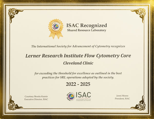
The Cleveland Clinic Research Flow Cytometry SRL is an ISAC-recognized facility.
The Flow Cytometry Core provides advanced capabilities for cell sorting, quantitative analysis of cells and extracellular vesicles, and single-cell transcriptomic profiling. The Core supports investigators through comprehensive services that include sample acquisition and analysis, high-speed cell sorting, and expert consultation for experimental design, data interpretation, and troubleshooting.
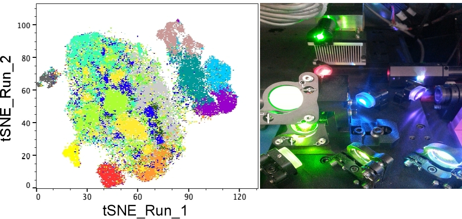
The facility houses a broad range of state-of-the-art instrumentation for conventional and spectral flow cytometry. Conventional flow cytometry platforms include a BD LSRFortessa equipped with five lasers and capable of 18-color, 20-parameter acquisition, and a BD FACSymphony A5 SE capable of high-dimensional 48-color, 50-parameter analysis using five lasers. For advanced immunophenotyping applications, the Core operates two spectral analyzers: a five-laser Cytek Aurora spectral analyzer equipped with a 96-well automated sampling system, and a six-laser Sony ID7000 spectral analyzer with automated sampling capabilities compatible with multiple well-plate formats.
For extracellular vesicle research, the Core provides a Particle Metrix ZetaView Quatt nanoparticle tracking analyzer equipped with four lasers, enabling high-resolution particle characterization and quantification.
The Core maintains full compliance with Biosafety Level 2 (BSL2) for cell sorting operations. Three high-speed cell sorters are housed within Class II biosafety cabinets to protect personnel and the environment from potentially aerosolized biological hazards. A BD FACSAria Fusion and Sony MA900 sorter are located within a dedicated 176-square-foot negative-pressure laboratory, while a Thermo Fihser Bigfoot high-speed cell sorter is housed in the Core's 1,300-square-foot main facility. All sorting platforms are equipped with aerosol management systems designed to evacuate aerosols generated within the sort chamber. In response to the COVID-19 pandemic, the Core implemented specialized standard operating procedures for handling clinical samples, based on recommendations from the International Society for Advancement of Cytometry and approved by the Cleveland Clinic Institutional Biosafety Committee. These procedures incorporate aerosol containment measures, respiratory protection for operators, and detailed emergency protocols for nozzle clogs and other aerosol-generating events.
Data acquisition is performed using SpectroFlo® software on the Cytek Aurora, Sony ID7000 software on the Sony spectral analyzer, Sasquatch software on the BigFoot, ZetaSphere for Zetaview, MA900 Sorting software for MA900 and BD FACSDiva software on the Fortessa, Symphony A5 SE and Fusion platforms. FACSDiva supports import and export of FCS 2.0, 3.0, and 3.1 file formats. List-mode data acquisition and storage allow investigators to analyze and present data in multiple formats tailored to their experimental needs. All data files are securely archived on Cleveland Clinic Research servers within designated laboratory folders, ensuring long-term accessibility for authorized users. The Core also provides FlowJo, terraFlow and ModFit software for advanced cytometric analysis.
To support single-cell genomics applications, the Core operates a Chromium X Controller for automated single-cell sample preparation and gene expression profiling. Services include evaluation of sample quality, optional cell sorting, GEM generation, thermal cycling, and cDNA amplification. Downstream library amplification, clean-up, and sequencing are performed in collaboration with the Genomics Core. To accommodate time-sensitive clinical specimens, the Core provides 24/7 support for fresh sample processing.
The validity, rigor, and operational excellence of the Flow Cytometry Core have been formally recognized by the International Society for Advancement of Cytometry through the ISAC Shared Resource Laboratory Recognition Program. The Core exceeded the threshold for excellence established under ISAC best practices for SRL operations. This recognition reflects compliance with current biosafety standards and rigorous performance metrics across Quality Assurance, Education and Training, Data Management, Laboratory Safety, Standard Operating Procedures, Operations, Staffing, and Infrastructure.
Acknowledgment Guidance
Acknowledgments and Authorship
All work performed by Cleveland Clinic Shared Laboratory Resources should be acknowledged or considered for co-authorship in scholarly publications, presentations, and posters. By acknowledging shared resource expertise and instrumentation, you play a critical role in supporting our mission to maintain access to cutting-edge technology and services on our campus into the future.
The Cleveland Clinic Shared Laboratory Resources participate in the Research Resource Identifier Initiative to promote transparency and reproducibility of scientific methods. Please include the unique Research Resource Identifier, RRID number, in the Methods or Acknowledgments sections of your manuscripts.
Example Acknowledgments:
- We acknowledge the Cleveland Clinic Research Flow Cytometry Core Facility (RRID:SCR_026460), especially [staff name], for their assistance with [technique/technology].
- This research was supported in part by Cleveland Clinic Research Flow Cytometry Shared Laboratory Resource (Cleveland, OH) (RRID:SCR_026460).
- This work was supported by NIH Shared Instrument S10 Grant S10OD025207-01A1 for the Sony ID700 Spectral Analyzer.