Research
The Yildirim Lab develops next-generation neurotechnologies and artificial intelligence frameworks to understand how brain circuits are altered across neurological disorders and cancer. Our work lies at the intersection of neuroscience, oncology, engineering, and data science, with a central focus on uncovering how disease reshapes neural dynamics, behavior, and tissue microenvironments across species.
A major emphasis of our research is cancer neuroscience, where we investigate how brain tumors such as glioblastoma disrupt neural circuit function, alter inter-regional communication, and drive changes in behavior. To address these questions, we build and deploy custom multimodal platforms that integrate large-scale optical imaging, electrophysiology, and high-resolution behavioral tracking—including pupil dynamics, orofacial activity, and locomotion. These approaches allow us to monitor the brain at unprecedented spatial and temporal scales during disease progression.
Complementing our experimental platforms, we develop AI-driven computational models to extract meaningful patterns from complex, high-dimensional datasets. Our machine learning tools enable the identification of noninvasive biomarkers of disease onset and progression, as well as predictive models that link neural activity to behavior and clinical phenotypes. By integrating multimodal data across scales—from single cells to whole-brain dynamics—we aim to define circuit-level signatures of disease.
Beyond discovery, our lab is advancing closed-loop neurotechnologies that combine real-time recording with targeted perturbation of neural circuits. These systems provide a framework to test whether restoring disrupted neural dynamics can improve functional outcomes, establishing neural circuit activity as both a biomarker and a therapeutic target.
In parallel, we extend these approaches across disease contexts, including neurodevelopmental disorders such as Autism Spectrum Disorder and cancer-related tissue remodeling in breast cancer. By leveraging cross-species models and translational datasets, our goal is to bridge fundamental neuroscience with precision medicine.
Ultimately, our research seeks to establish a new paradigm in which multimodal neurotechnology and AI enable mechanistic understanding, early detection, and circuit-level intervention in brain disorders and cancer.
Biography
Dr. Murat Yildirim holds a PhD degree from UT Austin and he received his B.Sc. and M.Sc. degrees from Middle East Technical University in Turkey, both in Mechanical Engineering. He performed his postdoctoral training at Picower Institute for Learning and Memory at the Massachusetts Institute of Technology where he developed custom-made optical systems to perform large scale optical recordings and perturbations in behaving mouse brain and cerebral organoids. His current research interests are developing next generation multiphoton systems to study the structure, function, and biomechanics of the brain with various biological systems and models spanning from cerebral organoids, to small animals, to humans through their interaction with femtosecond laser pulses for the diagnosis, characterization, and treatment of specific brain disorders to positively impact human mental health. He is the recipient of several prestigious awards such as a Pathway to Independence Award (K99/R00) from NIBIB. He is a member of SPIE, OSA, and SfN.
Education & Professional Highlights
Education & Fellowships
Ph.D. Mechanical Engineering – University of Texas at Austin
Austin, TX
2015
Postdoctoral Fellow – Picower Institute for Learning and Memory, Massachusetts Institute of Technology
Cambridge, MA
2022
Awards & Honors
National Institute of Biomedical Imaging and Bioengineering
Grant number: R00EB027706
National Institute of Biomedical Imaging and Bioengineering
Grant number: K99EB027706
Research
Research News
Our DeepFaceMouse Paper is Out (6/8/26)Excited to share that our latest paper, "DeepFaceMouse enables scalable prediction of large-scale brain activity from facial dynamics in mice," has been published in Cell Reports Methods by Cell Press
This work started with a simple but fundamental question:
Can facial dynamics serve as a noninvasive window into brain-wide neural activity?
To address this challenge, we developed DeepFaceMouse, a deep learning framework that accurately tracks mouse facial movements and predicts large-scale cortical activity from behavior alone.
Key innovations of this study:
🔹 High-precision tracking of eyelid, whisker, nose, and mouth movements using deep learning
🔹 Prediction of large-scale cortical activity from facial dynamics, enabling scalable brain–behavior analyses
🔹 Validation across multiple calcium indicators, including GCaMP6s, GCaMP6f, and jGCaMP8m, demonstrating robust performance across diverse neural imaging conditions
🔹 Generalization across multiple behavioral paradigms, including spontaneous behavior, virtual reality navigation, and social interaction assays
🔹 Successful application across distinct neurological disease models, including glioblastoma (GBM), autism spectrum disorder (ASD), and multiple sclerosis (MS)
🔹 Open-source software and user-friendly GUI designed for large-scale neuroscience studies
Our findings suggest that subtle facial movements contain rich information about internal brain states and that artificial intelligence can help bridge the gap between observable behavior and large-scale neural dynamics.
More broadly, DeepFaceMouse reflects our lab's vision of integrating neuroscience, engineering, and AI to develop scalable tools for understanding brain function in health and disease.
A huge thank you to first author Kemal Ozdemirli and all of my trainees, collaborators, and co-authors who contributed to this work. Also big thanks to Cleveland Clinic Research, American Brain Tumor Association Case Comprehensive Cancer Center The National Institutes of Health for their support!
📄 Paper: DeepFaceMouse enables scalable prediction of large-scale brain activity from facial dynamics in mice: Cell Reports Methods
Excited to share our new bioRxiv preprint from the Yildirim Lab:
“Deterministically Synchronized Widefield Imaging and Virtual Reality Platform for Multimodal Brain–Behavior Recording”
In this work, we developed a hardware-synchronized widefield calcium imaging and virtual reality (VR) platform that enables millisecond-precision alignment between:
• Brain-wide neural activity
• Pupil dynamics
• Orofacial behavior
• Locomotion
• Closed-loop VR environments
A major challenge in multimodal neuroscience experiments is that software-based synchronization can introduce latency, jitter, and timing drift — making it difficult to accurately study fast brain–behavior interactions. Our system addresses this by integrating all acquisition subsystems under a shared hardware-defined clock with deterministic synchronization and quantitatively validated timing performance.
Key features of the platform include:
🔹 Widefield cortical imaging with dual-wavelength hemodynamic correction
🔹 Simultaneous high-speed pupil and face recordings
🔹 Closed-loop VR integration with both ViRMEn and Blender
🔹 ~1.5 ms locomotion-to-VR update latency
🔹 Multimodal analyses including connectivity, motif extraction, spectral dynamics, and machine learning-based brain–behavior prediction
Using this framework, we demonstrate that mesoscale cortical activity during behavior is highly structured, low-dimensional, and strongly coupled to behavioral state variables such as locomotion and pupil dynamics.
This project represents a highly collaborative effort across neuroscience, engineering, optics, VR, and AI.
Great work led by another talented student Miguel Maldonado and we are grateful to everyone in the Yildirim Lab for their contributions.
Preprint: A Deterministically Synchronized Widefield Imaging and Virtual Reality Platform for Multimodal Brain–Behavior Recording | bioRxiv
Thanks to The National Institutes of Health Cleveland Clinic Research Case Comprehensive Cancer Center American Brain Tumor Association for their support!
Excited to share our new paper now available on bioRxiv: “NeuroPupil: A generalization-first framework for scalable and biologically informative cross-species pupillometry"
In this work, we developed NeuroPupil, a deep learning framework for high-throughput and precise pupil tracking across mice and humans. By systematically benchmarking training strategies, architectures, and aggregation methods, we identified pooled multi-subject training combined with optimized U-Net architectures as a key driver of robust and generalizable pupillometry.
Importantly, NeuroPupil was evaluated across diverse behavioral conditions, imaging resolutions, and disease contexts, including PTEN-associated autism spectrum disorder (ASD), glioblastoma (GBM), and cuprizone-induced multiple sclerosis (MS) mouse models, as well as human clinical datasets from pediatric strabismus patients. Across these datasets, NeuroPupil preserved biologically meaningful pupil dynamics that improved prediction of distributed cortical activity and enhanced classification of disease-associated pupil signatures.
These findings highlight how subtle improvements in pupil boundary localization can substantially influence downstream neural decoding, brain-state estimation, and translational biomarker discovery.
This project represents a major collaborative effort at the intersection of neuroscience, AI, behavior, and neurotechnology, and reflects the incredible work of our students, trainees, and collaborators.
It is led by our talented student Kemal Ozdemirli and we are very grateful to everyone in the lab and our collaborators who contributed to this work.
Link to the paper:
Thanks to The National Institutes of Health Case Comprehensive Cancer Center Cleveland Clinic Research American Brain Tumor Association for their support.
We are excited to share our latest collaborative study: “Integrated Collagen Architecture and Composition Improve Risk Stratification in Triple-Negative Breast Cancer.”
In this work, we combined multimodal histopathology, collagen imaging, and artificial intelligence–based computational analysis to investigate how extracellular matrix (ECM) organization influences disease progression in triple-negative breast cancer (TNBC).
Our findings show that quantitative collagen architecture and composition provide powerful prognostic information beyond conventional clinical markers, improving survival and recurrence risk stratification in TNBC patients. This study highlights the critical role of the tumor microenvironment in cancer progression and demonstrates the potential of AI-driven computational pathology for precision oncology.
This project represents a highly interdisciplinary collaboration across cancer biology, pathology, imaging, and machine learning.
We attended Optica Biophotonics Conference (05/04/26) I had a fantastic time at the Optica Biophotonics Congress this year!I’m especially proud to share that two of our lab’s abstracts were selected as finalists in the Optics in the Brain section — a great recognition of the team’s hard work and creativity.
A special shoutout to Tenesha Connor, who delivered an outstanding oral presentation, showcasing her side project and truly rocking the stage. It’s always rewarding to see trainees step into the spotlight with confidence and clarity.
Beyond the presentations, the conference was a great opportunity to reconnect with colleagues, spark new collaborations, and get inspired by a series of excellent plenary and invited talks pushing the boundaries of biophotonics and neuroscience.
Looking forward to the next one! Thanks for the support American Brain Tumor Association Case Comprehensive Cancer Center Cleveland Clinic Research
#Optica #Biophotonics #Neuroscience #AcademicResearch #Mentorship #Collaboration

I’m happy to share that our recent paper has been accepted (in principle) at Cell Reports Methods by Cell Press.
This project has been a true team effort, and I’m especially proud of Kemal Ozdemirli, our undergraduate researcher, who played a key role in driving this work forward with great dedication and curiosity.
A big thank you to all my lab members and collaborators for their contributions, support, and teamwork throughout this process. It’s been incredibly rewarding to see this project come together.
What makes this even more meaningful to me is that this work was fully developed here in Cleveland—from initial ideas to final results—highlighting the strength of our local scientific community.
Looking forward to sharing more once it’s out! Thanks for the support from American Brain Tumor Association Case Comprehensive Cancer Center Cleveland Clinic Research The National Institutes of Health

I am incredibly proud to share that my graduate student, Tenesha Connor, has been selected as an Emerging Graduate Student at the Cleveland Clinic Research.
Tenesha has been doing outstanding work in our lab, contributing to large-scale brain imaging and computational approaches to understand neural dynamics in health and disease. This recognition is a testament to her dedication, creativity, and scientific rigor.
It was a pleasure to attend the event together and celebrate this achievement with our entire lab team — moments like these truly highlight the importance of mentorship, teamwork, and a supportive research environment.
Congratulations, Tenesha — very well deserved, and excited for what’s ahead!

A great start to April! I had the opportunity to present my midterm seminar today, reflecting on the journey of building my lab at the Cleveland Clinic Research.
This milestone is truly a team effort. I am incredibly grateful to work alongside such a talented and dedicated group of trainees who are driving our work forward every day. Seeing how far we’ve come—from building new imaging systems to deploying deep learning models —has been deeply rewarding.Most importantly, this seminar reinforced where we are heading: toward scalable, translational tools that can bridge basic neuroscience and clinical impact. Excited for the next phase of growth, discovery, and collaboration.
Tenesha Connor received a best abstract award at MD Anderson International Cancer Neuroscience Symposium (02/20/26)
Congratulations our graduate student Tenesha who received a best abstract award at the Neuroplasm section at MD Anderson International Cancer Neuroscience symposium held on February 18-20, 2026.

Tenesha Connor presented her MSc research at the Cleveland State University today (02/05/26). Congratulations Tenesha for her hard work and MSc degree! We are excited to see her accomplishments in her PhD in our lab.

Dr. Yildirim chaired Human-Centered Health Innovation session at the Banu Onaral Symposium celebrating the legacy of Dr. Banu Onaral on January 30th, 2026. There were back-to-back talks today with Dr. Justin Lathia from totally different angles of glioblastoma (GBM) research- tumor biology meets brain dynamics and neurotechnology.

Proud of our amazing undergraduates for presenting their cutting-edge research at The Intersection at Case Western Reserve University! Their creativity, hard work, and dedication to science continue to inspire our whole lab. The future of neurophotonics, AI, and brain research is bright!

We are thrilled to announce that Yildirim lab at the Cleveland Clinic has been awarded a Discovery Grant from the American Brain Tumor Association (ABTA)! 🧠 We are deeply honored to be supported by such a prestigious organization and to join the ABTA Alumni Research Network (AARN). This award will help us advance our research on how glioblastoma disrupts brain networks and how targeted circuit modulation can restore function and improve outcomes.
Grateful to the ABTA for recognizing and supporting our work in this emerging field of cancer neuroscience — and to our incredible lab members and collaborators for their dedication and creativity in pushing this research forward.

The past two days at the Annual Retreat were truly inspiring- filled with insightful talks, innovative science, and the vibrant energy of our Cleveland cancer research community. We are incredibly proud of our students for their outstanding achievements:
🎓 Kemal Ozdemirli, whose abstract was selected among the Top 3 presentations — an exceptional recognition for an undergraduate researcher.🎓 Frederick Bell and 🎓 Tenesha Connor, who both received Best Poster Presentation Awards for their impressive work
These moments remind us why mentorship and discovery are at the heart of academic life. Watching our students grow, innovate, and represent our lab with excellence is one of the most rewarding parts of being a PI. Grateful to the Case CCC community for such a well-organized and inspiring event — and to our incredible team for their hard work, curiosity, and dedication.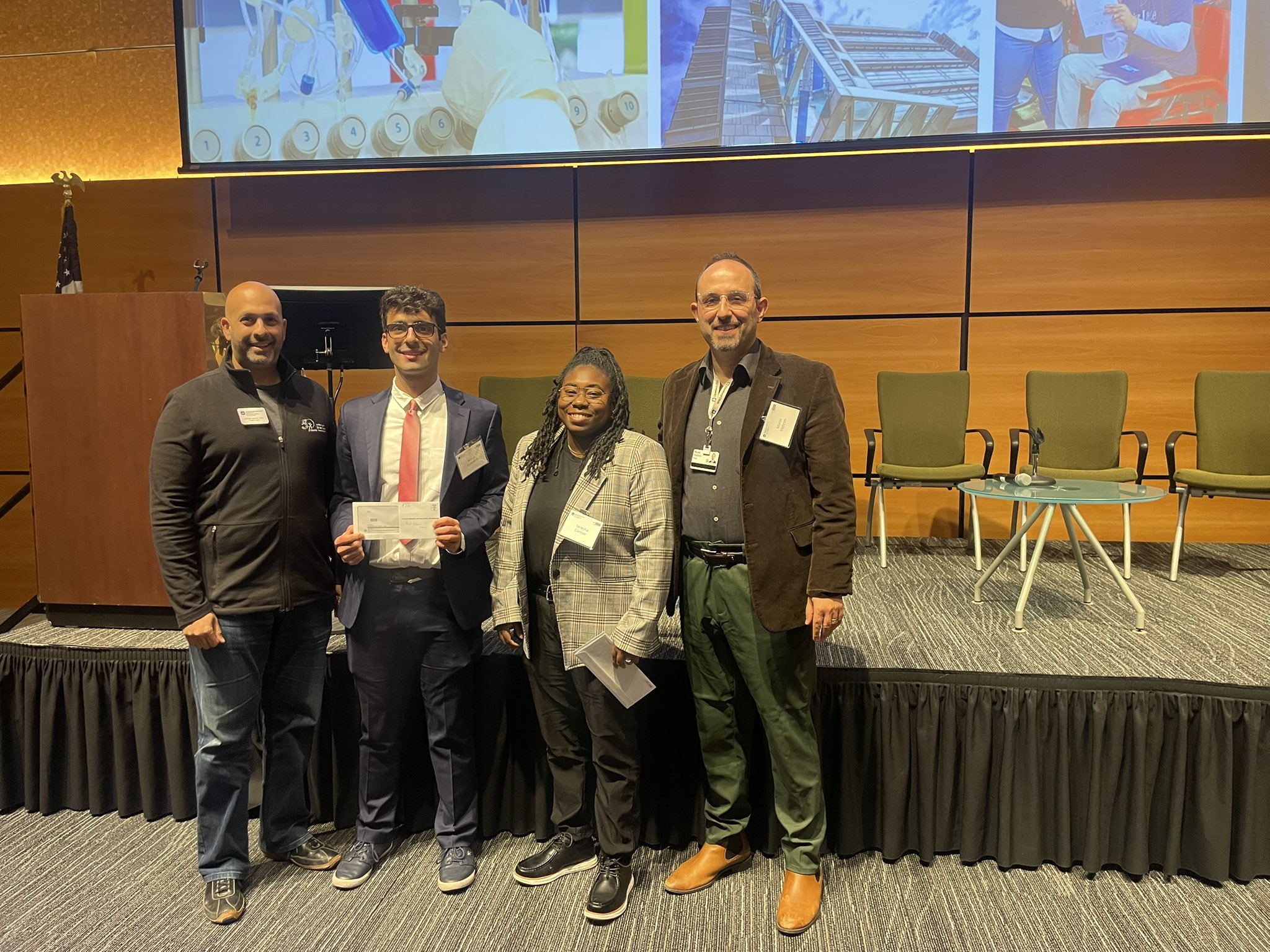 Celebrating Excellence at the Kent State Brain Health Research Symposium (10/20/25)
Our lab had an exciting and productive day at the Kent State University Brain Health Research Symposium, where we presented multiple posters highlighting our latest advances in neurophotonics, computational neuroscience, and brain-behavior analysis across neurological disorders.
We are especially proud of our graduate student Tenesha Connor, who was recognized with one of the Best Graduate Student Poster Awards for her outstanding work. This achievement reflects her dedication, creativity, and strong commitment to impactful brain health research. We are grateful to our collaborators and colleagues for inspiring discussions, valuable feedback, and continued support as we push the boundaries of neuroscience and work toward new discoveries that improve patient care. Congratulations to Tenesha and our entire team — onward to more breakthroughs! Our new paper is online! (06/19/25)
We’re excited to share our latest work: DeepFace, an ultrafast and highly precise deep learning tool for orofacial tracking and brain-state decoding in mice. From thousands of videos to high-throughput analysis pipelines, DeepFace is already transforming preclinical neuroscience. Here is the paper link.

Our stellar graduate student received two best poster awards at Cleveland State University! (04/24/25)
So proud of our first-year PhD student, Tenesha, for winning TWO poster awards today at the 8th Annual Thomas Bell Lecture at Cleveland State University — Best Immunity & Therapeutics Poster AND Best Overall Poster! 🎉🏆

Our undergraduate students presented their research at CWRU Research Day (04/23/25)
A truly proud PI moment today! 🎉Two of our incredible undergraduate students presented their research at the Intersections – Case Western Reserve University Undergraduate Research Day. One of their projects was highlighted in Case Daily, and the other student was recently awarded the Provost Summer Undergraduate Research Grant (PSURG) to continue their work this summer. It's been amazing to watch their growth as young scientists in our lab at the Cleveland Clinic Lerner Research Institute, as they’ve taken on complex challenges at the intersection of neuroscience, engineering, and computer science.I’m so proud of what they’ve accomplished—and excited for what’s ahead. The future is bright! ✨


New paper on Quantum Tensor Decomposition is online (02/20/25)
Tensor decomposition has emerged as a powerful framework for feature extraction in multi-modal biomedical data. In this review, we present a comprehensive analysis of tensor decomposition methods such as Tucker, CANDECOMP/PARAFAC, spiked tensor decomposition, etc. and their diverse applications across biomedical domains such as imaging, multi-omics, and spatial transcriptomics. To systematically investigate the literature, we applied a topic modeling-based approach that identifies and groups distinct thematic sub-areas in biomedicine where tensor decomposition has been used, thereby revealing key trends and research directions. We evaluated challenges related to the scalability of latent spaces along with obtaining the optimal rank of the tensor, which often hinder the extraction of meaningful features from increasingly large and complex datasets. Additionally, we discuss recent advances in quantum algorithms for tensor decomposition, exploring how quantum computing can be leveraged to address these challenges. Our study includes a preliminary resource estimation analysis for quantum computing platforms and examines the feasibility of implementing quantum-enhanced tensor decomposition methods on near-term quantum devices. Collectively, this review not only synthesizes current applications and challenges of tensor decomposition in biomedical analyses but also outlines promising quantum computing strategies to enhance its impact on deriving actionable insights from complex biomedical data. Here is the paper.

Tenesha's new paper got the cover page! (01/26/25)
Excited to share a major milestone for my graduate student! Their review paper on quantum dots (QDs) for advancing multiphoton microscopy (MPM) has been featured on the cover page of Photonics! This paper highlights how QDs are transforming deep brain imaging by overcoming challenges like light scattering, absorption, and tissue damage. With their high quantum yields, tunable emission spectra, and photostability, QDs enable deeper, clearer imaging and open new doors for functional imaging of neural activity and hemodynamic responses. Even more exciting is the potential for QDs in drug delivery and therapies, paving the way for innovative treatments for brain tumors and neurological disorders. I’m incredibly proud of my student’s dedication and hard work that made this achievement possible. 🎓👏 Check out the cover and full paper here

Our collaborative paper is on Biorxiv! (11/11/24)
Established methods for imaging the living mammalian brain have, to date, taken the brain’s optical properties as fixed; we here demonstrate that it is possible to modify the optical properties of the brain itself to significantly enhance at-depth imaging while preserving native physiology. Using a small amount of any of several biocompatible materials to raise the refractive index of solutions superfusing the brain prior to imaging, we could increase several-fold the signals from the deepest cells normally visible and, under both one-photon and two-photon imaging, visualize cells previously too dim to see. The enhancement was observed for both anatomical and functional fluorescent reporters across a broad range of emission wavelengths. Importantly, visual tuning properties of cortical neurons in awake mice, and electrophysiological properties of neurons assessed ex vivo, were not altered by this procedure. Paper link

Our graduate student is accepted to Cold Spring Harbor Imaging Course! (05/11/24)
Big congratulations to our stellar graduate student, Tenesha, for being accepted to attend
Cold Spring Harbor Laboratories 'Imaging Structure and Function in the Nervous System' course! Your hard work and dedication are truly paying off. Here's to the beginning of an exciting journey!

MS walk was great (05/04/24)
We attended MS walk today at Cleveland State University organized by National Multiple Sclerosis Society. We are not only raising awareness about Multiple Sclerosis but also witnessing firsthand how our lab's research might bring hope and change to patients' lives.

Solar Eclipse was amazing! (04/13/24)
Today, our lab had the incredible opportunity to witness a solar eclipse! It was truly a once-in-a-lifetime experience, sparking awe and inspiration among our team. Witnessing such a rare celestial event together strengthened our bond and fueled our passion for exploration!

New review paper on multiphoton imaging of cerebral and retinal organoids is published (03/05/24)
Congratulations to our postdoc Emre whose review paper on "Applications of multiphoton microscopy in imaging in cerebral and retinal organoids" is published in Frontiers in Neuroscience. Paper link

Happy New Year and Holidays! (12/24/23)
Happy Holidays and New Year from YildirimLab! We are very excited with our new discoveries that The New Year brings:)

Our undergraduate students, Kemal and Shreya, presented their work on the Undergraduate Student Research Day at Case Western University. Keep up your good work!

 New review paper is online (11/27/23)
Our review paper on "Advances in Ultrafast Lasers for Multiphoton Microscopy in Neuroscience" is online. Paper link is here.
 Congratulations to our undergraduate student for receiving a poster award! (10/27/23)
Our undergraduate student received a poster award at the Kent State Brain Conference. Congratulations Kemal, keep up the good work :)

We are so excited to organize Sculpted Light in the Brain 2024 in France (09/14/23)
We are SUPER excited to announce Sculpted Light in the Brain (SLB2024) which will take place in beautiful city of PARIS from 19-21 June 2024 at Cité Internationale Universitaire de Paris. More details to follow soon (shaping up to be our best yet).

Our collaborative work on behavioral neuroscience is just published in PlosOne Computational Biology (09/01/23)
In reversal learning tasks, the behavior of humans and animals is often assumed to be uniform within single experimental sessions to facilitate data analysis and model fitting. However, behavior of agents can display substantial variability in single experimental sessions, as they execute different blocks of trials with different transition dynamics. Here, we observed that in a deterministic reversal learning task, mice display noisy and sub-optimal choice transitions even at the expert stages of learning. We investigated two sources of the sub-optimality in the behavior. First, we found that mice exhibit a high lapse rate during task execution, as they reverted to unrewarded directions after choice transitions. Second, we unexpectedly found that a majority of mice did not execute a uniform strategy, but rather mixed between several behavioral modes with different transition dynamics. We quantified the use of such mixtures with a state-space model, block Hidden Markov Model (block HMM), to dissociate the mixtures of dynamic choice transitions in individual blocks of trials. Additionally, we found that blockHMM transition modes in rodent behavior can be accounted for by two different types of behavioral algorithms, model-free or inference-based learning, that might be used to solve the task. Combining these approaches, we found that mice used a mixture of both exploratory, model-free strategies and deterministic, inference-based behavior in the task, explaining their overall noisy choice sequences. Together, our combined computational approach highlights intrinsic sources of noise in rodent reversal learning behavior and provides a richer description of behavior than conventional techniques, while uncovering the hidden states that underlie the block-by-block transitions. Paper link is here.

Our collaborative work with Harvard and MIT is just published in Light: Science and Applications (08/15/23)
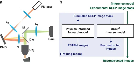
Limited throughput is a key challenge in in vivo deep tissue imaging using nonlinear optical microscopy. Point scanning multiphoton microscopy, the current gold standard, is slow especially compared to the widefield imaging modalities used for optically cleared or thin specimens. We recently introduced “De-scattering with Excitation Patterning” or “DEEP” as a widefield alternative to point-scanning geometries. Using patterned multiphoton excitation, DEEP encodes spatial information inside tissue before scattering. However, to de-scatter at typical depths, hundreds of such patterned excitations were needed. In this work, we present DEEP2, a deep learning-based model that can de-scatter images from just tens of patterned excitations instead of hundreds. Consequently, we improve DEEP’s throughput by almost an order of magnitude. We demonstrate our method in multiple numerical and experimental imaging studies, including in vivo cortical vasculature imaging up to 4 scattering lengths deep in live mice. Paper link is here.
Dr. Yildirim attended Sunposium and COSYNE conferences (04/01/23)
Dr. Yildirim attended back-to-back two conferences. First one is the Sunposium Conference held in West Palm Beach and the second one is COSYNE Conference held in Montreal.


First social outing (03/28/23)
We had our first social outing and our choice was bowling. We enjoyed celebrating our accomplishments in this event. Let's see what the next celebration is.

Our new lab logo is ready (02/01/23)
We have been working on designing a new lab logo. Now, it is ready to share with everyone. It represents that our hope is endless and our work never ends :)

Our abstract is accepted at Optica Biophotonics Conference (01/27/23)
We are so excited to present our work as an oral presentation at Optica Biophotonics Conference: Optics in the Life Sciences which will be held in Vancouver.

Our abstract is accepted at COSYNE 23 (01/27/23)
Our abstract is accepted as a poster presentation at COSYNE 23. We are delighted to join this prestigious conference.

Dr. Yildirim gave an invited talk at the Mayo Clinic Science of Medicine Ground Rounds (10/30/22)
Dr. Yildirim gave an invited talk entitled as " Of mice and men: Spatiotemporally-targeted high resolution brain imaging and optogenetics in health and disease" in the Mayo Clinic Science of Medicine Ground Rounds.

Dr. Yildirim was invited to talk at "Becoming a Faculty 101" Panel Discussion (09/23/22)
Dr. Yildirim was invited to share his experience on moving from his dependent to independent career at "Becoming a Faculty 101" Panel Discussion organized by Inet NYC. Inet NYC is a international scholar community providing support and professional guidance for all international graduate students and research fellows in STEM fields, that are or will be affiliated to any institution in the NYC area, to expand their skill set and career development opportunities.

New paper is published in Journal of Biomedical Optics (09/02/22)
We are so glad to publish this paper in collaboration with researchers at UT Austin. In this paper, we showed proof-of-concept application of creating subepithelial voids, injecting and localizing biomaterial for the treatment of vocal fold scarring. The paper link is here.

Our lab will be open on 09/01/22
Our lab will open its doors at the Neuroscience Department at Lerner Research Institute (LRI) in the Cleveland Clinic. We are looking for motivated technicians, graduate students, and postdocs to join our lab. We will be a multi-disciplinary group composed of neuroscientists, engineers, and medical doctors. If you are interested in making new discoveries in the brain through developing world-class technologies and working with world-class colleagues and collaborators, please check our open positions and lab policy/diversity pages.

Our Team
Selected Publications
2026Kemal Ozdemirli, Tenesha Connor, Kaleb Kim, Ersan Alp Unlu, Macit Emre Lacin, Miguel Maldonado, Frederick Bell, Thiago Peixoto Leal, Caglar Oksuz, Anthony Sloan, Nicholas Sarn, Anthony Chomyk, Bruce Trapp, Justin D. Lathia, Ignacio Mata, Charis Eng, Murat Yildirim (2026) We are excited to share that our latest paper, "DeepFaceMouse enables scalable prediction of large-scale brain activity from facial dynamics in mice," has been published in Cell Reports Methods by Cell Press
This work started with a simple but fundamental question:
Can facial dynamics serve as a noninvasive window into brain-wide neural activity?
To address this challenge, we developed DeepFaceMouse, a deep learning framework that accurately tracks mouse facial movements and predicts large-scale cortical activity from behavior alone.
Key innovations of this study:
🔹 High-precision tracking of eyelid, whisker, nose, and mouth movements using deep learning
🔹 Prediction of large-scale cortical activity from facial dynamics, enabling scalable brain–behavior analyses
🔹 Validation across multiple calcium indicators, including GCaMP6s, GCaMP6f, and jGCaMP8m, demonstrating robust performance across diverse neural imaging conditions
🔹 Generalization across multiple behavioral paradigms, including spontaneous behavior, virtual reality navigation, and social interaction assays
🔹 Successful application across distinct neurological disease models, including glioblastoma (GBM), autism spectrum disorder (ASD), and multiple sclerosis (MS)
🔹 Open-source software and user-friendly GUI designed for large-scale neuroscience studies
Our findings suggest that subtle facial movements contain rich information about internal brain states and that artificial intelligence can help bridge the gap between observable behavior and large-scale neural dynamics.
More broadly, DeepFaceMouse reflects our lab's vision of integrating neuroscience, engineering, and AI to develop scalable tools for understanding brain function in health and disease.
A huge thank you to first author Kemal Ozdemirli and all of my trainees, collaborators, and co-authors who contributed to this work. Also big thanks to Cleveland Clinic Research, American Brain Tumor Association Case Comprehensive Cancer Center The National Institutes of Health for their support!
📄 Paper: DeepFaceMouse enables scalable prediction of large-scale brain activity from facial dynamics in mice: Cell Reports Methods
Miguel Maldonado, Omer Faruk Dinc, Macit Emre Lacin, Tenesha Connor, Frederic Bell, Berfin Dinc, Kemal Ozdemirli, Murat Yildirim. bioRxiv (2026)
We are excited to share our new bioRxiv preprint from the Yildirim Lab:
“Deterministically Synchronized Widefield Imaging and Virtual Reality Platform for Multimodal Brain–Behavior Recording”
In this work, we developed a hardware-synchronized widefield calcium imaging and virtual reality (VR) platform that enables millisecond-precision alignment between:
• Brain-wide neural activity
• Pupil dynamics
• Orofacial behavior
• Locomotion
• Closed-loop VR environments
A major challenge in multimodal neuroscience experiments is that software-based synchronization can introduce latency, jitter, and timing drift — making it difficult to accurately study fast brain–behavior interactions. Our system addresses this by integrating all acquisition subsystems under a shared hardware-defined clock with deterministic synchronization and quantitatively validated timing performance.
Key features of the platform include:
🔹 Widefield cortical imaging with dual-wavelength hemodynamic correction
🔹 Simultaneous high-speed pupil and face recordings
🔹 Closed-loop VR integration with both ViRMEn and Blender
🔹 ~1.5 ms locomotion-to-VR update latency
🔹 Multimodal analyses including connectivity, motif extraction, spectral dynamics, and machine learning-based brain–behavior prediction
Using this framework, we demonstrate that mesoscale cortical activity during behavior is highly structured, low-dimensional, and strongly coupled to behavioral state variables such as locomotion and pupil dynamics.
This project represents a highly collaborative effort across neuroscience, engineering, optics, VR, and AI.
Great work led by another talented student Miguel Maldonado and we are grateful to everyone in the Yildirim Lab for their contributions.
We are excited to share our new paper now available on bioRxiv: “NeuroPupil: A generalization-first framework for scalable and biologically informative cross-species pupillometry"
We are excited to share our new paper now available on bioRxiv: “NeuroPupil: A generalization-first framework for scalable and biologically informative cross-species pupillometry"
In this work, we developed NeuroPupil, a deep learning framework for high-throughput and precise pupil tracking across mice and humans. By systematically benchmarking training strategies, architectures, and aggregation methods, we identified pooled multi-subject training combined with optimized U-Net architectures as a key driver of robust and generalizable pupillometry.
Importantly, NeuroPupil was evaluated across diverse behavioral conditions, imaging resolutions, and disease contexts, including PTEN-associated autism spectrum disorder (ASD), glioblastoma (GBM), and cuprizone-induced multiple sclerosis (MS) mouse models, as well as human clinical datasets from pediatric strabismus patients. Across these datasets, NeuroPupil preserved biologically meaningful pupil dynamics that improved prediction of distributed cortical activity and enhanced classification of disease-associated pupil signatures.
These findings highlight how subtle improvements in pupil boundary localization can substantially influence downstream neural decoding, brain-state estimation, and translational biomarker discovery.
This project represents a major collaborative effort at the intersection of neuroscience, AI, behavior, and neurotechnology, and reflects the incredible work of our students, trainees, and collaborators.
We are excited to share our latest collaborative study: “Integrated Collagen Architecture and Composition Improve Risk Stratification in Triple-Negative Breast Cancer.”
In this work, we combined multimodal histopathology, collagen imaging, and artificial intelligence–based computational analysis to investigate how extracellular matrix (ECM) organization influences disease progression in triple-negative breast cancer (TNBC).
Our findings show that quantitative collagen architecture and composition provide powerful prognostic information beyond conventional clinical markers, improving survival and recurrence risk stratification in TNBC patients. This study highlights the critical role of the tumor microenvironment in cancer progression and demonstrates the potential of AI-driven computational pathology for precision oncology.
This project represents a highly interdisciplinary collaboration across cancer biology, pathology, imaging, and machine learning.
2025 Kemal Ozdemirli, Tenesha Connor, Kaleb Kim, Macit Emre Lacin, Miguel Maldonado, Thiago Peixoto Leal, Caglar Oksuz, Ignacio Mata, Murat Yildirim. bioRxiv (2025)We present DeepFace, a next-generation facial analysis pipeline that enhances orofacial tracking and cortical activity prediction in mice. Rather than replacing existing tools, DeepFace builds upon DeepLabCut and Facemap to address scalability bottlenecks and improve behavioral quantification. It offers high precision, keypoint customization, and robust performance across GCaMP6s, GCaMP6f, and jGCaMP8m lines. With scalable batch processing and high-performance computing compatibility, DeepFace enables high-throughput brain-behavior analysis in large-scale preclinical neuroscience
Tensor decomposition has emerged as a powerful framework for feature extraction in multi-modal biomedical data. In this review, we present a comprehensive analysis of tensor decomposition methods such as Tucker, CANDECOMP/PARAFAC, spiked tensor decomposition, etc. and their diverse applications across biomedical domains such as imaging, multi-omics, and spatial transcriptomics. To systematically investigate the literature, we applied a topic modeling-based approach that identifies and groups distinct thematic sub-areas in biomedicine where tensor decomposition has been used, thereby revealing key trends and research directions. We evaluated challenges related to the scalability of latent spaces along with obtaining the optimal rank of the tensor, which often hinder the extraction of meaningful features from increasingly large and complex datasets. Additionally, we discuss recent advances in quantum algorithms for tensor decomposition, exploring how quantum computing can be leveraged to address these challenges. Our study includes a preliminary resource estimation analysis for quantum computing platforms and examines the feasibility of implementing quantum-enhanced tensor decomposition methods on near-term quantum devices. Collectively, this review not only synthesizes current applications and challenges of tensor decomposition in biomedical analyses but also outlines promising quantum computing strategies to enhance its impact on deriving actionable insights from complex biomedical data.
Tenesha Connor,Hemal Weerasinghe,Justin Lathia,Clemens Burda,Murat Yildirim,"Advances in Deep Brain Imaging with Quantum Dots: Structural, Functional, and Disease Specific Roles" Photonics (2025)Quantum dots (QDs) have emerged as promising tools in advancing multiphoton microscopy (MPM) for deep brain imaging, addressing long-standing challenges in resolution, penetration depth, and light–tissue interactions. MPM, which relies on nonlinear photon absorption, enables fluorescence imaging within defined volumes, effectively reducing background noise and photobleaching. However, achieving greater depths remains limited by light scattering and absorption, compounded by the need for balanced laser power to avoid tissue damage. QDs, nanoscale semiconductor particles with unique optical properties, offer substantial advantages over traditional fluorophores, including high quantum yields, large absorption cross-sections, superior photostability, and tunable emission spectra. These properties enhance signal to background ratio at increased depths and reduce scattering effects, making QDs ideal for imaging subcortical regions like the hippocampus without extensive microscope modifications. Studies have demonstrated the capability of QDs to achieve imaging depths up to 2100 μm, far exceeding that of conventional fluorophores. Beyond structural imaging, QDs facilitate functional imaging applications, such as high-resolution tracking of hemodynamic responses and neural activity, supporting investigations of neuronal dynamics and blood flow in vivo. Their stability enables long-term, targeted drug delivery and photodynamic therapy, presenting potential therapeutic applications in treating brain tumors, Alzheimer’s disease, and traumatic brain injury. This review highlights the impact of QDs on MPM, their effectiveness in overcoming light attenuation in deep tissue, and their expanding role in diagnosing and treating neurological disorders, positioning them as transformative agents for both brain imaging and intervention.
2024 Tenesha Connor,Hemal Weerasinghe,Justin Lathia,Clemens Burda,Murat Yildirim,"Advances in Deep Brain Imaging with Quantum Dots: Structural, Functional, and Disease Specific Roles" Preprints (2024)Quantum dots (QDs) have emerged as promising tools in advancing multiphoton microscopy (MPM) for deep brain imaging, addressing long-standing challenges in resolution, penetration depth, and light-tissue interactions. MPM, which relies on nonlinear photon absorption, enables fluorescence imaging within defined volumes, effectively reducing background noise and photobleaching. However, achieving greater depths remains limited by light scattering and absorption, compounded by the need for balanced laser power to avoid tissue damage. QDs, nanoscale semiconductor particles with unique optical properties, offer substantial advantages over traditional fluorophores, including high quantum yields, large absorption cross-sections, superior photostability, and tunable emission spectra. These properties enhance signal to background ratio at increased depths and reduce scattering effects, making QDs ideal for imaging subcortical regions like the hippocampus without extensive microscope modifications. Studies have demonstrated the capability of QDs to achieve imaging depths up to 2100 μm, far exceeding that of conventional fluorophores. Beyond structural imaging, QDs facilitate functional imaging applications, such as high-resolution tracking of hemodynamic responses and neural activity, supporting investigations of neuronal dynamics and blood flow in vivo. Their stability enables long-term, targeted drug delivery and photodynamic therapy, presenting potential therapeutic applications in treating brain tumors, Alzheimer’s disease, and traumatic brain injury. This review highlights the impact of QDs on MPM, their effectiveness in overcoming light attenuation in deep tissue, and their expanding role in diagnosing and treating neurological disorders, positioning them as transformative agents for both brain imaging and intervention.
Giovanni Talei Franzesi, Ishan Gupta, Ming Hu, Kiryl Piatkevich, Murat Yildirim, Jian-Ping Zhao, Minho Eom, Seungjae Han, Demian Park, Himashi Andaraarachchi, Zhaohan Li, Jesse Greenhagen, Amirul Muhammad Islam, Parth Vashishtha, Zahid Yaqoob, Nikita Pak, Alexander D Wissner-Gross, Daniel A. Martin-Alarcon, Jonathan J. Veinot, Peter T. C. So, Uwe Kortshagen, Young-Gyu Yoon, Mriganka Sur, Edward S. Boyden "In Vivo Optical Clearing of Mammalian Brain" Biorxiv(2024)Established methods for imaging the living mammalian brain have, to date, taken the brain’s optical properties as fixed; we here demonstrate that it is possible to modify the optical properties of the brain itself to significantly enhance at-depth imaging while preserving native physiology. Using a small amount of any of several biocompatible materials to raise the refractive index of solutions superfusing the brain prior to imaging, we could increase several-fold the signals from the deepest cells normally visible and, under both one-photon and two-photon imaging, visualize cells previously too dim to see. The enhancement was observed for both anatomical and functional fluorescent reporters across a broad range of emission wavelengths. Importantly, visual tuning properties of cortical neurons in awake mice, and electrophysiological properties of neurons assessed ex vivo, were not altered by this procedure.
Macit Emre Lacin, and Murat Yildirim "Applications of multiphoton microscopy in imaging cerebral and retinal organoids", Frontiers in Neuroscience (2024)Cerebral organoids, self-organizing structures with increased cellular diversity and longevity, have addressed shortcomings in mimicking human brain complexity and architecture. However, imaging intact organoids poses challenges due to size, cellular density, and light-scattering properties. Traditional one-photon microscopy faces limitations in resolution and contrast, especially for deep regions. Here, we first discuss the fundamentals of multiphoton microscopy (MPM) as a promising alternative, leveraging non-linear fluorophore excitation and longer wavelengths for improved imaging of live cerebral organoids. Then, we review recent applications of MPM in studying morphogenesis and differentiation, emphasizing its potential for overcoming limitations associated with other imaging techniques. Furthermore, our paper underscores the crucial role of cerebral organoids in providing insights into human-specific neurodevelopmental processes and neurological disorders, addressing the scarcity of human brain tissue for translational neuroscience. Ultimately, we envision using multimodal multiphoton microscopy for longitudinal imaging of intact cerebral organoids, propelling advancements in our understanding of neurodevelopment and related disorders.
2023 Thulasi Srinivasan, and Murat Yildirim "Advances in Ultrafast Fiber Lasers for Multiphoton Microscopy in Neuroscience", Photonics (2023)Multiphoton microscopy (MPM) has emerged as a vital tool in neuroscience, enabling deeper imaging with a broader field of view, as well as faster and sub-cellular resolution. Recent innovations in ultrafast fiber laser technology have revolutionized MPM applications in living brains, offering advantages like cost-effectiveness and user-friendliness. In this review, we explore the progress in ultrafast fiber laser technology, focusing on its integration into MPM for neuroscience research. We also examine the utility of femtosecond fiber lasers in fluorescence and label-free two- and three-photon microscopy applications within the field. Furthermore, we delve into future possibilities, including next-generation fiber laser designs, novel laser characteristics, and their potential for achieving high spatial and temporal resolution imaging. We also discuss the integration of fiber lasers with implanted microscopes, opening doors for clinical and fundamental neuroscience investigations.
Navodini Wijethilake, Mithunjha Anandakumar, Cheng Zheng, Peter T.C. So, Murat Yildirim, Dushan N. Wadduwage"DEEP2: Deep learning powered de-scattering with Excitation Patterning", Light: Science and Applications (2023)Limited throughput is a key challenge in in vivo deep tissue imaging using nonlinear optical microscopy. Point scanning multiphoton microscopy, the current gold standard, is slow especially compared to the widefield imaging modalities used for optically cleared or thin specimens. We recently introduced “De-scattering with Excitation Patterning” or “DEEP” as a widefield alternative to point-scanning geometries. Using patterned multiphoton excitation, DEEP encodes spatial information inside tissue before scattering. However, to de-scatter at typical depths, hundreds of such patterned excitations were needed. In this work, we present DEEP2, a deep learning-based model that can de-scatter images from just tens of patterned excitations instead of hundreds. Consequently, we improve DEEP’s throughput by almost an order of magnitude. We demonstrate our method in multiple numerical and experimental imaging studies, including in vivo cortical vasculature imaging up to 4 scattering lengths deep in live mice.
Nhat Minh Le, Murat Yildirim, Yizhi Wang, Hiroki Sugihara, Mehrdad Jazayeri, Mriganka Sur "Mixtures of strategies underlie rodent behavior during reversal learning", Plos ONe Computational Biology (2023)In reversal learning tasks, the behavior of humans and animals is often assumed to be uniform within single experimental sessions to facilitate data analysis and model fitting. However, behavior of agents can display substantial variability in single experimental sessions, as they execute different blocks of trials with different transition dynamics. Here, we observed that in a deterministic reversal learning task, mice display noisy and sub-optimal choice transitions even at the expert stages of learning. We investigated two sources of the sub-optimality in the behavior. First, we found that mice exhibit a high lapse rate during task execution, as they reverted to unrewarded directions after choice transitions. Second, we unexpectedly found that a majority of mice did not execute a uniform strategy, but rather mixed between several behavioral modes with different transition dynamics. We quantified the use of such mixtures with a state-space model, block Hidden Markov Model (block HMM), to dissociate the mixtures of dynamic choice transitions in individual blocks of trials. Additionally, we found that blockHMM transition modes in rodent behavior can be accounted for by two different types of behavioral algorithms, model-free or inference-based learning, that might be used to solve the task. Combining these approaches, we found that mice used a mixture of both exploratory, model-free strategies and deterministic, inference-based behavior in the task, explaining their overall noisy choice sequences. Together, our combined computational approach highlights intrinsic sources of noise in rodent reversal learning behavior and provides a richer description of behavior than conventional techniques, while uncovering the hidden states that underlie the block-by-block transitions.
2022 Navodini Wijethilake, Mithunjha Anandakumar, Cheng Zheng, Peter T.C. So, Murat Yildirim, Dushan N. Wadduwage"DEEP2: Deep learning powered de-scattering with Excitation Patterning", Arxiv. org (2022)
Limited throughput is a key challenge in in-vivo deep-tissue imaging using nonlinear optical microscopy. Point scanning multiphoton microscopy, the current gold standard, is slow especially compared to the wide-field imaging modalities used for optically cleared or thin specimens. We recently introduced 'De-scattering with Excitation Patterning or DEEP', as a widefield alternative to point-scanning geometries. Using patterned multiphoton excitation, DEEP encodes spatial information inside tissue before scattering. However, to de-scatter at typical depths, hundreds of such patterned excitations are needed. In this work, we present DEEP2, a deep learning based model, that can de-scatter images from just tens of patterned excitations instead of hundreds. Consequently, we improve DEEP's throughput by almost an order of magnitude. We demonstrate our method in multiple numerical and physical experiments including in-vivo cortical vasculature imaging up to four scattering lengths deep, in alive mice.
Ilan Gabay, Kaushik Subramanian, Liam Andrus, Andrew DuPlissis, Murat Yildirim, Adela Ben-Yakar"In vivo hamster cheek pouch subepithelial ablation, biomaterial injection, and localization: pilot study", Journal of Biomedical Optics(2022)Significance: The creation of subepithelial voids within scarred vocal folds via ultrafast laser ablation may help in localization of injectable biomaterials toward a clinically viable therapy for vocal fold scarring.
Aim: We aim to prove that subepithelial voids can be created in a live animal model and that the ablation process does not engender additional scar formation. We demonstrate localization and long-term retention of an injectable biomaterial within subepithelial voids.
Approach: A benchtop nonlinear microscope was used to create subepithelial voids within healthy and scarred cheek pouches of four Syrian hamsters. A model biomaterial, polyethylene glycol tagged with rhodamine dye, was then injected into these voids using a custom injection setup. Follow-up imaging studies at 1- and 2-week time points were performed using the same benchtop nonlinear microscope. Subsequent histology assessed void morphology and biomaterial retention.
Results: Focused ultrashort pulses can be used to create large subepithelial voids in vivo. Our analysis suggests that the ablation process does not introduce any scar formation. Moreover, these studies indicate localization, and, more importantly, long-term retention of the model biomaterial injected into these voids. Both nonlinear microscopy and histological examination indicate the presence of biomaterial-filled voids in healthy and scarred cheek pouches 2 weeks postoperation.
Conclusions: We successfully demonstrated subepithelial void formation, biomaterial injection, and biomaterial retention in a live animal model. This pilot study is an important step toward clinical acceptance of a new type of therapy for vocal fold scarring. Future long-term studies on large animals will utilize a miniaturized surgical probe to further assess the clinical viability of such a therapy.
Navodini Wijethilake, Cheng Zheng, Jong K Park, Murat Yildirim, Udith Haputhanthri, Peter TC So, Dushan N Wadduwage"DEEP learning powered De-scattering with Excitation Patterning (DEEP) for high-throughput wide-field multiphoton microscopy", SPIE(2022)Multiphoton microscopy is the gold standard for deep tissue fluorescence imaging. Long wavelengths enable hundreds of microns deep penetration of excitation light, but the emission fluorescence at shorter wavelengths encounters scattering before detection. While not being an issue for point scanning geometries, for wide-field geometries emission light scattering degrades the image quality. In this work, we use temporally focused pattered excitations to spatially encode image information before emission light scattering. Upon detection, images are reconstructed computationally by solving a linear inverse problem. We further improve our results by learning inverse solvers and optimal patterns through physics-based deep learning.
Nhat Minh Le, Murat Yildirim, Yizhi Wang, Hiroki Sugihara, Mehrdad Jazayeri, and Mriganka Sur. "Mixture of Learning Strategies Underlies Rodent Behavior in Dynamic Foraging." bioRxiv (2022).In volatile foraging environments, animals need to adapt their learning in accordance with the uncertainty of the environment and knowledge of the hidden structure of the world. In these contexts, previous studies have distinguished between two types of strategies, model-free learning, where reward values are updated locally based on external feedback signals, and inference-based learning, where an internal model of the world is used to make optimal inferences about the current state of the environment. Distinguishing between these strategies during the dynamic foraging behavioral paradigm has been a challenging problem for studies of reward-guided decisions, due to the diversity in behavior of model-free and inference-based agents, as well as the complexities that arise when animals mix between these types of strategies. Here, we developed two solutions that jointly tackle these problems. First, we identified four key behavioral features that together benchmark the switching dynamics of agents in response to a change in reward contingency. We performed computational simulations to systematically measure these features for a large ensemble of model-free and inference-based agents, uncovering an organized structure of behavioral choices where observed behavior can be reliably classified into one of six distinct regimes in the two respective parameter spaces. Second, to address the challenge that arises when animals use multiple strategies within single sessions, we developed a novel state-space method, block Hidden Markov Model (blockHMM), to infer switches in discrete latent states that govern the choice sequences across blocks of trials. Our results revealed a remarkable degree of mixing between different strategies even in expert animals, such that model-free and inference-based learning modes often co-existed within single sessions. Together, these results invite a re-evaluation of the stationarity of behavior during dynamic foraging, provide a comprehensive set of tools to characterize the evolution of learning strategies, and form the basis of understanding neural circuits involved in different modes of behavior within this domain.
Murat Yildirim, Chloe Delepine, Danielle Feldman, Vincent Pham, Stephanie Chou, Jacque Pak Kan Ip, Alexi Nott et al. "Label-free three-photon imaging of intact human cerebral organoids: tracking early events in brain development and deficits in Rett Syndrome." Elife (2022).Human cerebral organoids are unique in their development of progenitor-rich zones akin to ventricular zones from which neuronal progenitors differentiate and migrate radially. Analyses of cerebral organoids thus far have been performed in sectioned tissue or in superficial layers due to their high scattering properties. Here, we demonstrate label-free three-photon imaging of whole, uncleared intact organoids (∼2 mm depth) to assess early events of early human brain development. Optimizing a custom-made three-photon microscope to image intact cerebral organoids generated from Rett Syndrome patients, we show defects in the ventricular zone volumetric structure of mutant organoids compared to isogenic control organoids. Long-term imaging live organoids reveals that shorter migration distances and slower migration speeds of mutant radially migrating neurons are associated with more tortuous trajectories. Our label-free imaging system constitutes a particularly useful platform for tracking normal and abnormal development in individual organoids, as well as for screening therapeutic molecules via intact organoid imaging.
Careers
Postdoctoral Scholar
Computational Neuroscientist for Analyzing and Modeling Large Scale Recordings and Manipulations in Health and Disease
We are looking for a postdoctoral scholar who will analyze in vivo large-scale optogenetics and imaging dataset with behaving preclinical models in health and disease. With these analysis pipelines, we would like to understand how neural circuits generate patterned activity that gives rise to complex behaviors and phenotypes in health and disease states. It is a fully-funded and three-years long postdoctoral position. Its duration can be extended further.
Who we are: We are highly-motivated multi-disciplinary research group focuses on cracking brain complexity in the lens of engineering, optics, photonics, behavioral and computational neuroscience for basic and translational neuroscience questions. We are developing cutting edge microscopes which allows us to record and manipulate neuronal activity in various spatial and temporal scales in health and disease. Please visit our website (yildirimlab.org).
Who we are looking for: We are looking for a curious computational neuroscientist who is interested in working in a team-oriented environment and interested in making new discoveries in the basic and translational neuroscience. These positions are ideal for someone with a desire to develop skillsets with analyzing large scale recordings and manipulations and dissect neural circuits responsible for preclinical behavior in health and disease states.
Responsibilities: It is required to have a doctoral degree in Neuroscience, Engineering, or related disciplines, and to have expertise on one of these following topics: modeling preclinical behavior such as reinforcement learning and spatial learning, generalized linear model (GLM) for analyzing neural recordings, machine learning such as recurrent neural networks (RNN) for preclinical behavior and neural recordings, and Hidden Markov Modeling (HMM) for analyzing preclinical behavior. It is also required to have experience with coding (MATLAB, R, or Phyton), excellent organizational, analytical, and oral and written communication skills, ability to analyze data and present it in a format suitable for publication, self-motivation, and ability to function effectively in a teamoriented environment.
Preferences: It is preferred to have a doctoral degree in Neuroscience, and to have an expertise in GLM, HMM, and RNN.
Interested candidates should submit CV, cover letter, and contact information of two-three references to Dr. Murat Yildirim, [email protected].
Research News

Tool uses artificial intelligence to track facial feature movement and predict brain activity across 20–30 regions.

Building on promising early data, a Cleveland Clinic team secured funding to jump start their project on how we assess glioblastoma’s progression and potential injury to the brain.

Research aims to extend observations of reversal learning in preclinical models to human neurological disorders

An ongoing collaboration between Dr. Murat Yildirim and colleagues at Harvard and MIT aims to make it easier to track neurological activity deep within brain tissue.